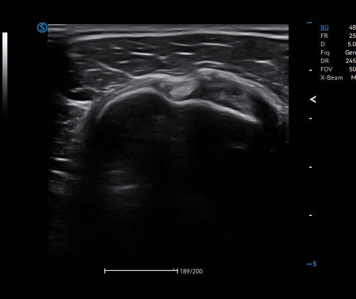
Color Doppler


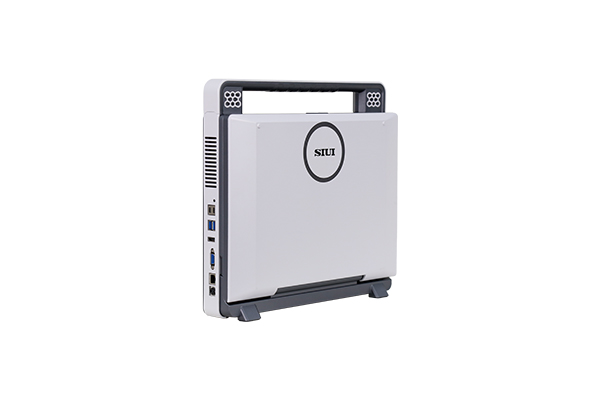
Apogee C1
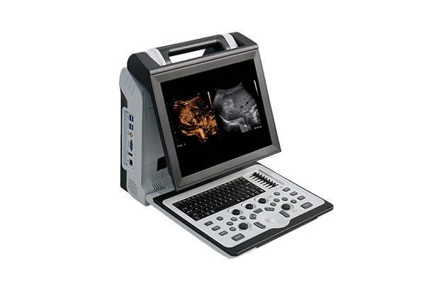
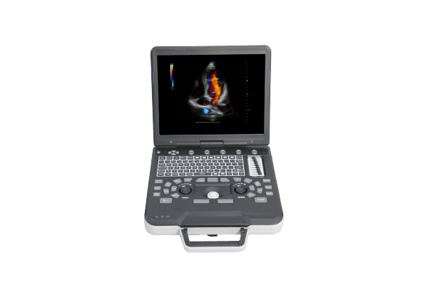
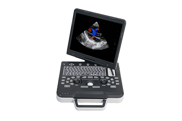
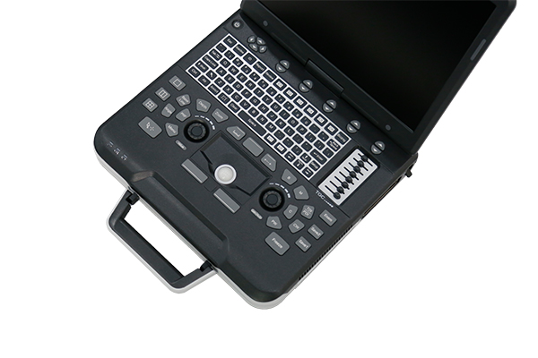
Apogee C1 is an entry-level ultrasound system, tailored for primary care.
It brings comprehensive fundamental exams to community clinics and health centers, and with its high portability, reaches patients even at home.
Features
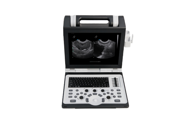
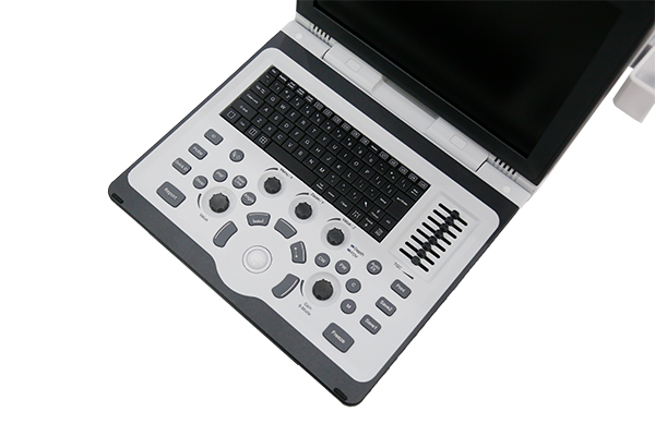
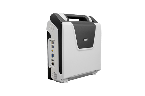
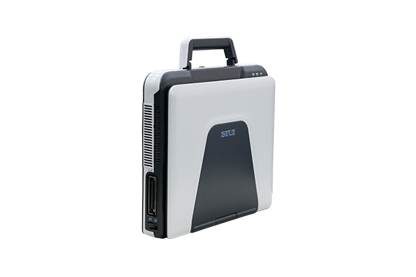
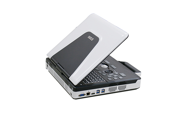
Elegant Outlook Design
• 14.6-inch adjustable monitor
• 3 probe connectors
• Built-in battery supports up to 2 hours of operation
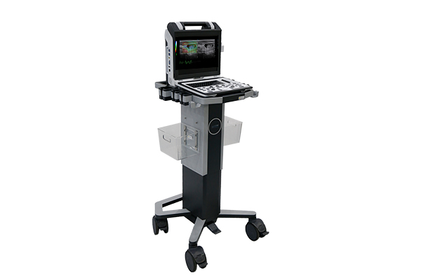
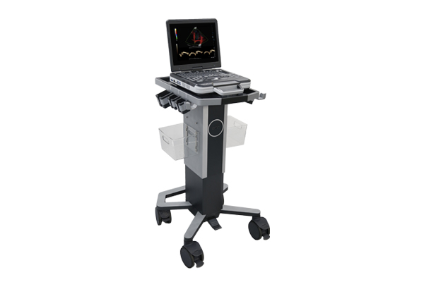
Compatible with Trolley
• Adjustable height
• 6 stable probe holders
• Omni-directional wheels
Function
Click on images to view more
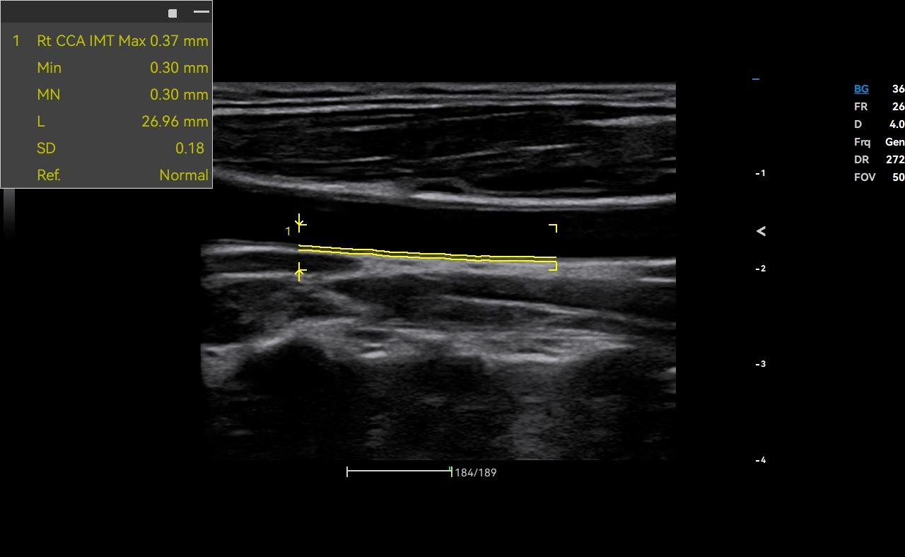
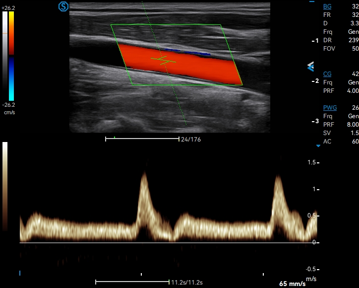
Auto IMT automatically measures the intima-media thickness of the carotid artery for early diagnosis of cardiovascular diseases.

When identifying a lesion, doctors can activate Auto Aspect Ratio by one keystroke to acquire essential quantitative results for growth orientation evaluation.

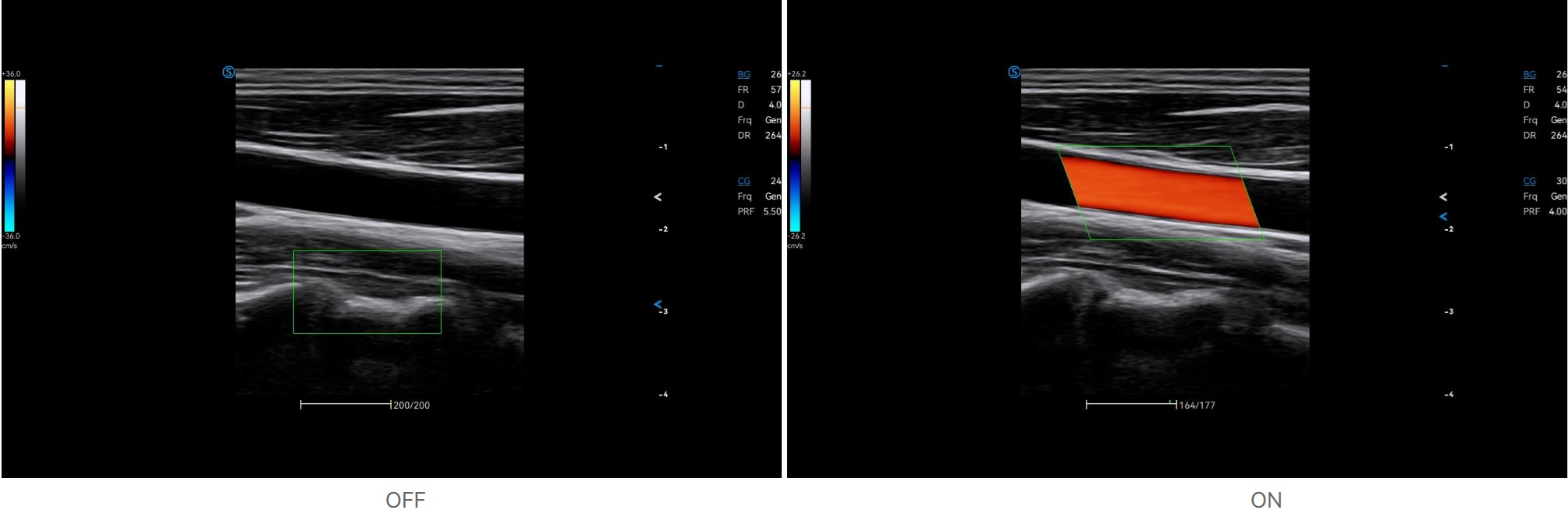
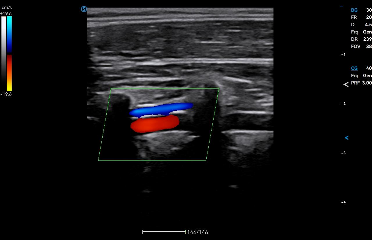
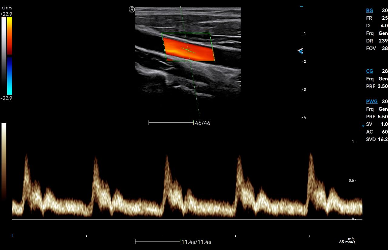
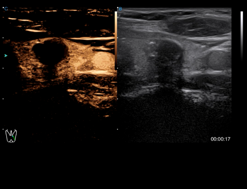
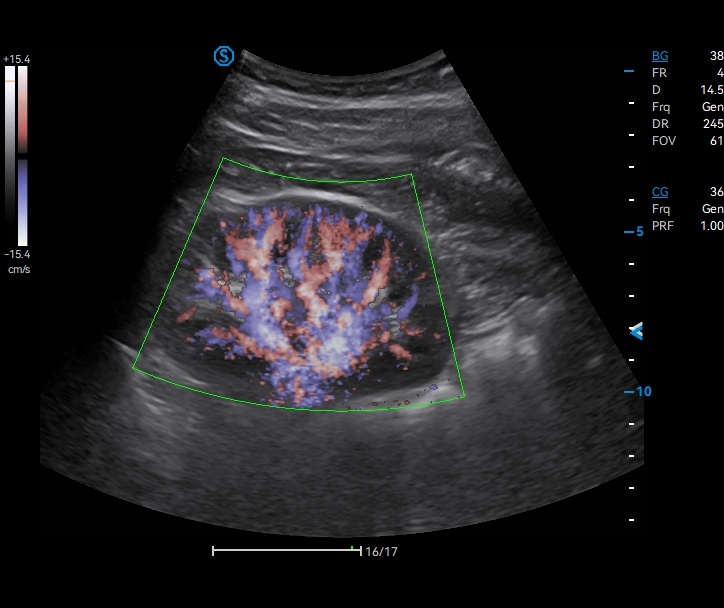
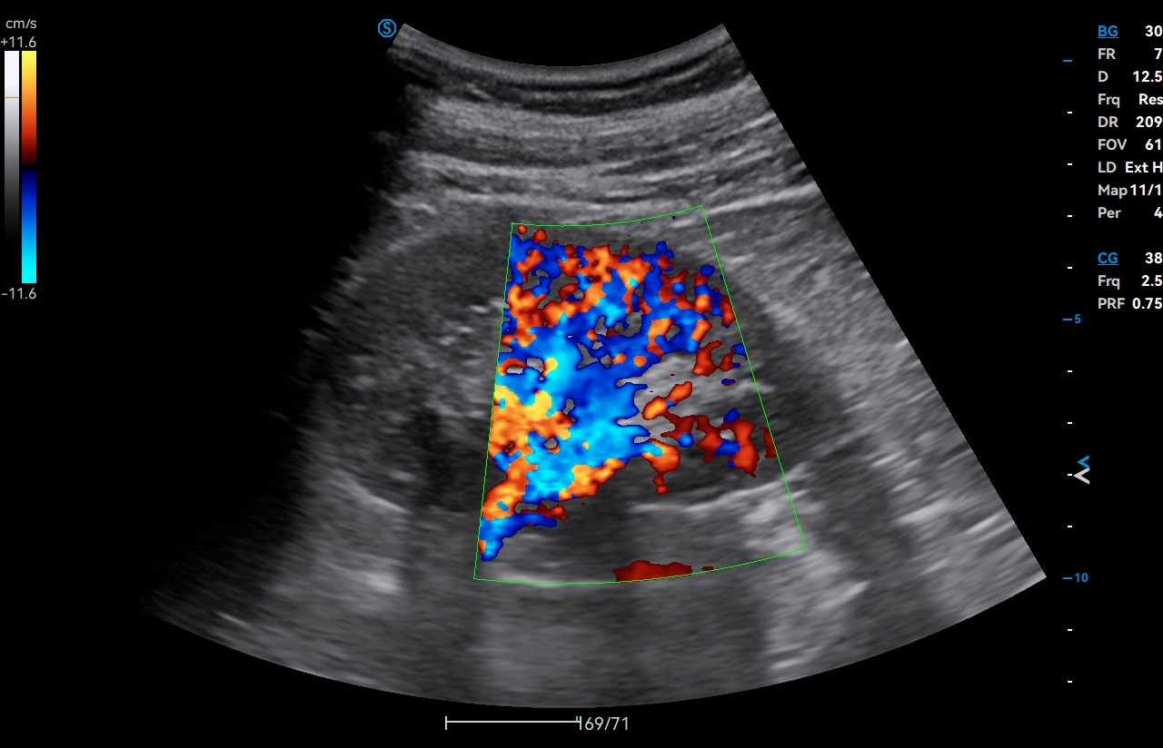
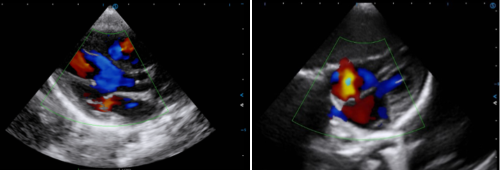
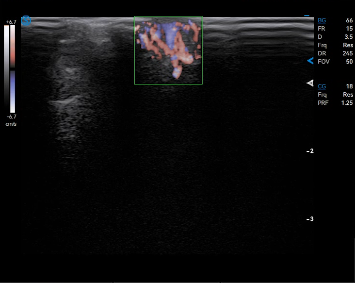
VS Flow is highly sensitive to low velocity blood flow signal, while Micro Flow sensitively captures the micro blood flow signal.


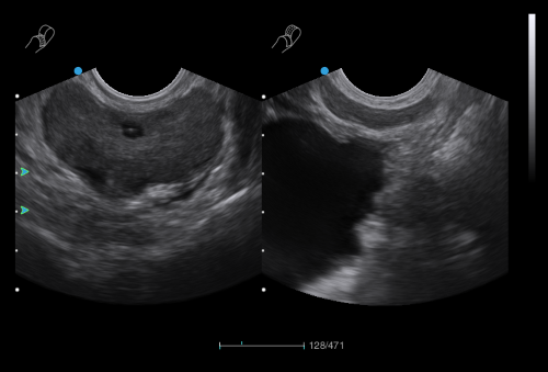
Auto FLC automatically detects, envelops, and measures the biggest follicle. This function would help in tracking follicle growth for infertility treatment.

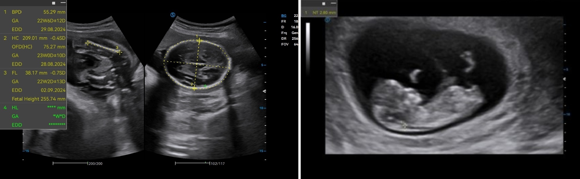
Auto OB automatically measures the common fetal biometric results (e.g. HC, BPD, FL, etc.) in standard sections.




IBUS
- Details
- Category: Color Doppler
 Click on images to enlarge


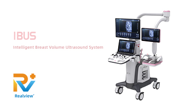
IBUS
Superb Versatility in Women’s and Children’s Healthcare
The IBUS series comprises IBUS 60 and IBUS 30. IBUS is the intelligent breast volume diagnostic system that provides integrated solutions for both women and children. The specialized breast probe, standardized scanning process, and standardized diagnosis procedure guided by BI-RADS significantly enhance diagnostic accuracy for breast examinations. The cutting-edge application solutions in OB / GYN / pediatrics aim at elevating the quality of healthcare for women and children.
Features
Outlook Design
- High-resolution monitor
- Dual touch screens
- Specialized breast probe and 3 common probe connectors
- Large-sized breast probe for efficient scanning

- Patented coupling film for enhanced patient comfort
- Low damping articulated arm with wide range of adjustment

Standardized & Customizable Workflow
- Standardized process avoids missed diagnosis
- Customizable scanning sections adapt to specific cases
IBUS vs Traditional Ultrasound
- Standardized screening minimizes reliance on experience and skill
- Enhance tumor detection, better reveal the morphology of minute lesions
- Free 3D imaging displays coronal, transverse, and sagittal planes, reducing the misdiagnosis rate
- Accurate spatial stereo positioning, highly consistent with surgical fields
- Field-of-view is over 3 times larger than traditional 2D probe to capture large lesions completely
IBUS vs Mammography
- High repeatability with no radiation dosage
- More accurate and convenient for diagnosing solid-cystic tumors
- Higher detection rate of tumors in dense breast
- Display breast with volume ultrasonic tomography
- Easily acquire images of coronal, transverse, and sagittal planes
- Accurate spatial stereo positioning, highly consistent with surgical fields
IBUS vs MRI
- Shorter examination time and higher diagnostic efficiency
- Harmless and applicable to all people
- Cost-effective
Functions
Full Volume Imaging and Coronal Section with High Clinical Value
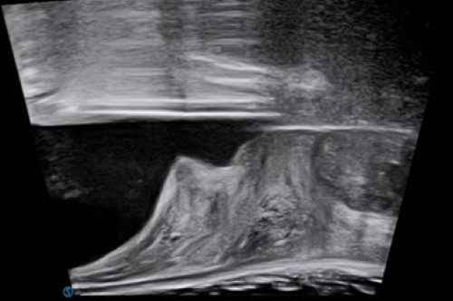
IBUS acquires volume data from multiple sections to provide abundant information. The coronal section intuitively shows the anatomical information of breast tissue in a supine position during operation, which helps surgeon to perform more accurate surgical planning.

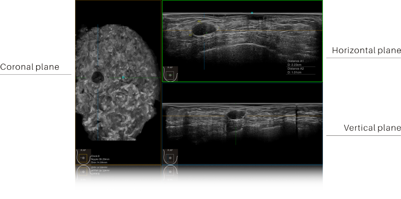
Intelligent Breast Solutions on the Workstation
Automatic lesion detection powered by AI identifies suspected area and traces the lesion from multiple sections. Quantitative coordinate position and graphic indication will be displayed for precise lesion localization.

Lesion stereo images are generated from volume data rendering. It provides intuitive and detailed information for better observation and lesion localization.
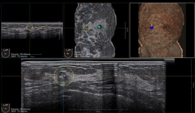
Lesion correlation correlates the information obtained from different scanning positions and sections to avoid misdiagnosis.

Auto OB automatically measures the common fetal biometric results (HC, BPD, FL, etc.) in standard sections.

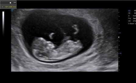
Auto NT automatically recognizes and measures the thickness of the fetal nuchal translucency.
Lumi 4D enables the adjustment of light source angle to support real-time static stereo imaging of the fetus.

Pediatrics
Craniocerebral Ultrasound Tomography (Infant) automatically identifies the 12 standard craniocerebral sections of the pediatric brain through volume data

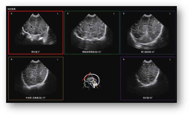
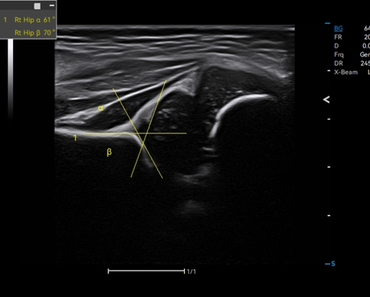
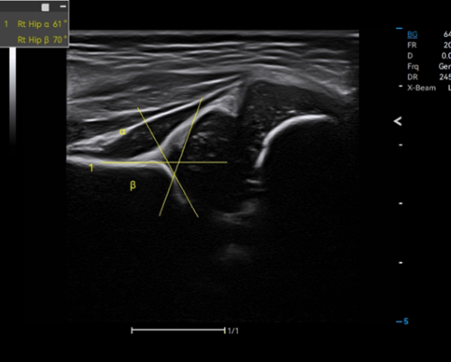
Auto Hip identifies the baseline, tracks the top line of bone and cartilage, and measures crucial results for hip dysplasia evaluation.



 Click on images to enlarge